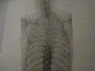
1. The flow pictures
2. The blood pool pictures
3. The pictures you take 3 hours after the other ones.
The flow pictures is just the Tc-MDP being whisked around the body. What you are expecting once you see hot flow is that there will be a hot bone scan. If there's not, then think of cellulitis.
However, the main usefulness of the flows is to tell you if the process is acute or subacute/chronic - a normal flow will mean the latter.
The blood pool pictures happen because of the Tc-MDP you inject, not all of it is able to stick to the bone crystals. About half does, and the other half is just sitting in all of the capillaries in the body - this is what makes the soft tissues look hot.
A few hours later, all of the stuff that was unable to bind to the bone is peed out.
This is why on the delayed pictures, the kidneys - which are now full of Tc-MDP - should look as hot as the lumbar vertebrae.
There is still about 5% of the dose you injected sitting in the capillaries, so the soft tissue never completely disappears on those delayed images.
However, what you should NOT be seeing is anything else apart from bone and urinary system.
If you do then the causes are:
If see THYROID and STOMACH - free pertechnetate (maybe there wasn't enough stannous in that vial to reduce all the pertechnetate).
If see LIVER and SPLEEN - alumina has leaked out of the generator and has led to clumps/colloid of alumina-Tc forming.
If see LUNG - aluminium has leaked out of the generator and has not formed clumps, but has bound to the Tc and caused it to get stuck in the lungs.
The first thing to look at on the delayed scan is for EXTRA-OSSEUS UPTAKE.
This means you wil not miss ascites or pleural effusion, which occur because the Tc-MDP leaks out of the blood in areas where the vessels are leaky.
Know that breast uptake does not equal malignancy, because fibroadenoma and mastitis will cause it too.
If you see dots of uptake in the soft tissues then this may be:
- injection sites or skin contamination (e,g, urine) or contamination of the camera screen
- free calcium from tissue destruction (inc.radiotherapy)
- free calcium from conditions that cause calcium deposition (hyperPTH, sarcoidosis). However, just like kidney stones only form in acidic urine, these will only deposit in acidic areas - fundus, kidneys, lungs.
- ossification
- amyloid (because amyloid precursor protein has a high calcium content)
The next thing to look for is to see if you can see the kidneys.
If you can't then it is either a SUPERSCAN OR there is renal failure OR the bones are so packed with iron that there is no space for the MDP and therefore the whole picture looks fuzzy.
It is easy to tell the difference between the two:
- in renal failure, the whole body looks like a blood pool image and the bones are fuzzy.

Then, finally, you look at the skeleton.
First, look for cold spots.
A classic cold spot to be aware of is avascular necrosis.
Then, look for hot spots.
The principle here is that not all hotspots are abnormal.
It is normal for all of the S's to be hot:
SKULL - hyperostosis frontalis
SPHENOID - a butterfly shaped bone that sits between the eyes where it forms part of the skull base
SCARF - the neck at the site of the laryngotracheal cartilage, especially if there is a lordosis
STERNOMANUBRIAL JOINT and the STERNAL OSSIFICATION CENTRE
SIDE - all of the bones on either side of the joints in an arm or a leg that is dominant
SACROLIACS
SPINE - you see vertical linear activity due to insertion of the erector spinae.
...and in a child, the EPIPHYSES
...and in a neonate get crap pictures because nothing is calcified.
Thinking about this in a differnet way:-
Normal scans show:
- hot sternomanubrial joint and hot around the shoulder joints
- sinuses are hot
- ilia are hot on the anterior view because they're closest to the camera.
- sacro-iliac joints hot on the posterior view because they're closest to the camera.
- midline of spine hottest on the posterior view because it's closest to the camera.
- skeleton looks like they have a tonsure on their head
- sternum hotter than the ribs on the anterior view because it's closest to the camera
 So, the normal skeleton looks like a monk with a peg on his nose, a tonsure and a big cross on his chest and epaulets on his shoulders.
SO, IF IT'S NOT ONE OF THESE S SPOTS THEN IT'S NOT NORMAL.
THE NEXT QUESTION IS: IS THE HOT SPOT A WORRY SPOT?
1. Spots near joints:
The rule is that if it's on both sides of the joint, then it's not a worry spot.
There are 2 exceptions to this rule:
i) If you are worried about septic arthritis, then it is still a worry spot
ii) If it is in the sternum in a patient with ipsilateral breast cancer then it is a metastasis until proven otherwise.
2. Spots anywhere else:
It's simple. They are ALL worry spots.
So, the follow-on question is simple too because almost all of these spots will be either trauma or metastasis. That question is:- do they have a trauma pattern or a metastasis pattern?
Trauma pattern=
The third and last thing to account for on the three-phase bone scan that gives three mSv is to look for cold spots.
If you see one then this means pure osteoclast action - this is typically seen in plasmacytoma.
However, once the weakened bone fractures, osteoblasts move in and the area becomes hot.
The final thing is to take special views to get more of an idea about a hot spot. Typical examples are:
- if there is ?something in the pelvis, but the bladder overlies it, use a SQUAT view
- if there is something at the junction of the rib and scapular tip, use an ARMS UP view
- if there is ?something on the side curve of the ribs, use an OBLIQUE view.
Other things to know about bone scans:
- The radionuclide you use is Technetium bound to a diphosphonate, the latter being the bit that gets into the bone. How do you get this?
1. You milk the Mo-99 cow and out comes a radioactive milk called Pertechnetate (chemical symbol 99m-TcO4-)
2. If you then put this milk into a vial with a reducing agent called stannous then you get 99m-Tc).
3. If you add something else into the vial then the 99m-Tc will stick to that something else. For bone imaging, that something else is Diphosphonate.
This disphosphonate will get taken up by calcifying apatite and becomes a part of the bone. So, note that it is NOT osteoblasts that take up the radioactivity, but newly forming bone itself.
- The way that you create Tc-diphosphonate is that the Tc that comes off the generator is put into a vial that already has stuff in it. This stuff is stannous and diphosphonate. The stannous is there to change the Tc in such a way ("reduce it")
- You use a dose of 600MBq (20mCi), which gives an effective dose to the patient of 3milliSV - this being the dose that they would get from a year's worth of living in a world with sunshine and granite.
1. Bone scans are maps of osteoblast function, all that you can see is:
- hyperfunction, normal function or hypofunction.
In a fat person, everything looks hypo because it's so attenuated.
The major patterns of hyperfunction are due to FRACTURE, MALIGNANCY AND ARTHRITIS.
These three are differentiated thus:
FRACTURE is punctate for long bones and plate-like for vertebrae.

MALIGNANCY expands bone and travels along the bone for much longer than fracture.
ARTHRITIS occurs on the inner curve of the spine, and is hot because of osteophytes.

In the end, if you don't know, do as Nathan B. does, and order a plain X-ray of the region.
Other causes are due to:
- PAGETS if the bone is thicker than those around it and the hyperfunction begins at the joint end of a long bone or in the skull. So, this is like Graves disease in appearance - you get hyperfunction.
- OSTEOSARCOMA occurs in the metaphysis (i.e. almost the end) of long bones and in the pelvis. It metastasizes to the lungs.
- WEIGHT BEARING if all the bones in one leg are hyper compared to the other leg.
- RADIOTHERAPY causes cold bones because it has burnt out all the osteoblasts.
The differential for a cold bone is an infarct or metal.

- CELLULITIS if the blood pool scan shows a hot area but the 4 hour pictures show that the bone in that area is not hot.
The trouble with bone scans is that there are usually just so many hot spots to describe!
The best way to describe the bits that aren't important it is to say that "normal activity is also seen in the kidneys & bladder and degenerative changes in the hands, feet, spine and shoulders"
- a common misconception is that if an area is hotter then it is because it has more bone being formed in it than another area. Not the case!
The reason an area is hotter is because:
1. it has more Tc sitting in capillaries because there is a greater blood flow to the area. This is why

No comments:
Post a Comment