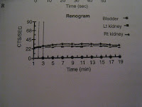
There are two curves:
1. The perfusion curve, which will show the amount of isotope in the aorta, and the amount of isotope in the kidneys over the first minute of imaging. What you should see is that the kidney lines meet the aorta line. If they don't then that means there is sluggish perfusion.
 This is what a normal perfusion scan looks like:
What does that mean? Well, it's like what happens in chronic PE - the rest of the arterioles grow thick and so there is reduced perfusion. Thus, in chronically sick kidneys, there is increased resistance to flow, and so you get reduced perfusion.
2. The clearance curve, shows the amount of tracer in the kidneys over the next 1/2 hour of imaging. It should reach a peak in the first few minutes and then decline. If it doesn't go down then that means that very little is being:
a. filtered through the glomerulus or secreted through the tubules if you are using MAG-3 as the isotope.
b. filtered if you are using DTPA as the isotope.

If there is still nothing in the bladder from one side then you give Frusemide (0.5mg/kg up to a dose of 40mg) to see if that washes the tracer down into the bladder.
So, the final 2 things to look at if you are seeing these renal failure curves are images, not curves.
1. Delayed image to make sure that there is no obstruction to outflow.
2. Amount of tracer in the soft tissues, because this tells you how high the creatinine is.
So, what if you just wanted to do everything off the pictures. Well you should see:
- perfusion occurring by 30 seconds
- nephrogram occurring by 1 minute
- collecting system outline by 5 mins
- nothing much except for the bladder to be seen by 20 mins
If, however, you want to do everything from the pictures only then this is a good example:
a. It doesn't show the perfusion images, but that part is easy to interpret. The question is - does perfusion occur at the same time as the aorta appears? If yes, then you say "perfusion is prompt". If not, then you say "perfusion is delayed".
b. "Uptake is seen on both sides", but is "not symmetrical" because of the photopaenic defect on the left. That's all that you can ever say about uptake.
c. Clearance is seen - you can say this if you see the tracer go from cortex into the calyceal system. And then you have to look at the last picture and compare it to the first one with clearance. If things are as black then as now, then "clearance is poor". Else, clearance is satisfactory.
d. Finally, look to see if the bladder is seen and both ureters are full outlined. If so, then "drainage is seen without obstruction".
That's it!
Other stuff to know about renal scans:
- the reason why MAG-3 is preferred is that it has that dual way of getting into the collecting system. Therefore you get crisper pictures in renal failure.
Examples of abnormal scans:
- Urinoma


No comments:
Post a Comment